Endochondral ossification is a developmental process of bone formation that occurs through a cartilage intermediate. It is a highly intricate event involving various cell types, such as bone cells, vessel cells, and cells from the bone marrow niche. At present, it is not possible to fully replicate endochondral ossification under controlled laboratory conditions. This process is essential for both bone development and repair following injury, and it also contributes to the progression of several debilitating conditions, including osteoarthritis.
In our lab, we have developed several in vivo models for bone formation using a cartilage intermediate created from differentiated MSCs. These constructs can be implanted directly under the skin of mice or be placed in a bovine bone ring to create a critical-sized defect in a semi-orthopedic model. Our goal is to leverage these models to advance bone tissue engineering, test innovative biomaterials, evaluate implant osseointegration, study vascularization, explore the immune response in bone defect healing, develop antibacterial therapies for implant-related infections, investigate osteonecrosis and marrow niche formation, and examine metastasis and leukemia.
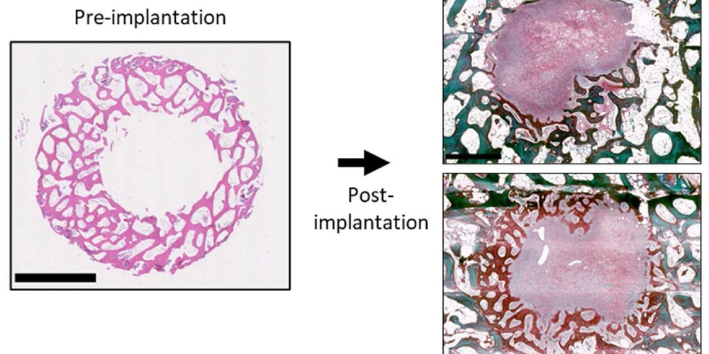
Image: Semi-orthotopic model for critical size bone defect healing [E. Andrés Sastre et al., Biomaterials 2021]
People
Andrea Lolli, Jietao Xu (Alumnus)
Barrett, H. E., Van der Heiden, K., Farrell, E., Gijsen, F. J. H., and Akyildiz, A. C. (2019)
J Biomech 87, 1-12
Leszczynska, A., O'Doherty, A., Farrell, E., Pindjakova, J., O'Brien, F. J., O'Brien, T., Barry, F., and Murphy, M. (2016)
Stem Cells 34, 913-923
Kenswil, K. J. G., Pisterzi, P., Sanchez-Duffhues, G., van Dijk, C., Lolli, A., Knuth, C., Vanchin, B., Jaramillo, A. C., Hoogenboezem, R. M., Sanders, M. A., Feyen, J., Cupedo, T., Costa, I. G., Li, R., Bindels, E. M. J., Lodder, K., Blom, B., Bos, P. K., Goumans, M. J., Ten Dijke, P., Farrell, E., Krenning, G., and Raaijmakers, M. (2021)
Cell Stem Cell 28, 653-670 e611
Andres Sastre, E., Maly, K., Zhu, M., Witte-Bouma, J., Trompet, D., Bohm, A. M., Brachvogel, B., van Nieuwenhoven, C. A., Maes, C., van Osch, G., Zaucke, F., and Farrell, E. (2021)
Bone 150, 115999